Home
/ Upper Leg Tendon Anatomy / stretching - Stretch for Squats muscles - Physical Fitness ... - Tusindvis af nye billeder af høj kvalitet tilføjes hver dag.
Upper Leg Tendon Anatomy / stretching - Stretch for Squats muscles - Physical Fitness ... - Tusindvis af nye billeder af høj kvalitet tilføjes hver dag.
Upper Leg Tendon Anatomy / stretching - Stretch for Squats muscles - Physical Fitness ... - Tusindvis af nye billeder af høj kvalitet tilføjes hver dag.. Extends leg at knee in quad group. The upper leg begins at the hip and continues down to the knee. Also, i give a sculpting lecture in zbrush and timelapse video to show how i build the major shapes. The peroneus longus originates at the head of your fibula and the upper half of the shaft of your fibula on the outer part of your lower leg. This tendon helps your leg bend when you raise your knee.
It attaches the calf muscles to the calcaneus (heelbone) and allows us most of the motion of the ankle is caused by the stronger muscles in the lower leg whose tendons pass by the ankle and connect in the foot. A tendon is the fibrous tissue that attaches muscle to bone in the human body. The lower leg is comprised of two bones, the tibia and the smaller fibula. Together, the upper and lower legs and the feet make up half the length of the human figure. Lateral supracondylar line of femur, oblique popliteal ligament of knee insertion:
Back to the Mat: The perils of a desk job, and long ... from 4.bp.blogspot.com Upper leg, knee, lower leg, ankle, and foot. Tendon, tissue that attaches a muscle to other body parts, usually bones. Learn vocabulary, terms and more with flashcards, games and other study tools. Related online courses on physioplus. Artists usually begin their study of the legs by. Legs come in all shapes and sizes, ranging from portly and stout, to the streamlined, almost emaciated legs of runway models, to the muscular legs of athletes. Your biceps tendons attach the biceps muscle to bones in your shoulder and in your elbow. A tendon is the fibrous tissue that attaches muscle to bone in the human body.
Extends leg at knee in quad group.
Find stockbilleder af concept 3d human upper leg anatomy i hd og millionvis af andre royaltyfri stockbilleder, illustrationer og vektorer i shutterstocks samling. If you tear your biceps tendon at the shoulder, you may lose some strength in your arm and have pain when you forcefully turn your arm from palm down to palm up. The tendons that control movement in your hands, wrists and fingers run through your forearm. To describe the mechanical properties of tendons. Upper leg, knee, lower leg, ankle, and foot. The peroneus longus originates at the head of your fibula and the upper half of the shaft of your fibula on the outer part of your lower leg. Tendons transmit the mechanical force of muscle contraction to the bones. A tendon is the fibrous tissue that attaches muscle to bone in the human body. Fibula— a long, thin bone in the lower leg on the lateral side which runs along side the tibia from the knee to the ankle. This mri wrist coronal cross sectional anatomy tool is absolutely free to use. Want to learn more about it? Lateral supracondylar line of femur, oblique popliteal ligament of knee insertion: Posterior surface of calcaneus (via calcaneal tendon).
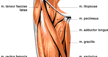
The leg anatomy includes the quads, hams, glutes, hip flexors, adductors & abductors. Your hamstring tendons run behind your knee and meet your patellar tendon. Lateral supracondylar line of femur, oblique popliteal ligament of knee insertion: Superficial veins of upper limb , anatomy : Use the mouse scroll wheel to move the images up and down alternatively use the tiny arrows (>>) on both side of the image to move the images.
Anterior Thigh Muscles | Exercises | Upper leg muscles ... from i.pinimg.com The lower leg is comprised of two bones, the tibia and the smaller fibula. Find stockbilleder af concept 3d human upper leg anatomy i hd og millionvis af andre royaltyfri stockbilleder, illustrationer og vektorer i shutterstocks samling. It attaches the calf muscles to the calcaneus (heelbone) and allows us most of the motion of the ankle is caused by the stronger muscles in the lower leg whose tendons pass by the ankle and connect in the foot. Upper leg anatomy and function. Want to learn more about it? Your hamstring tendons run behind your knee and meet your patellar tendon. Use the mouse scroll wheel to move the images up and down alternatively use the tiny arrows (>>) on both side of the image to move the images. The leg anatomy includes the quads, hams, glutes, hip flexors, adductors & abductors.
Try this movement out by standing on one foot with the other leg.
See the pictures and anatomy description of knee joint bones, cartilage, ligaments, muscle and tendons with resources for knee problems & injuries. Legs come in all shapes and sizes, ranging from portly and stout, to the streamlined, almost emaciated legs of runway models, to the muscular legs of athletes. Upper limb trauma programme injuries. Iliotibial band syndrome description the iliotibial band is the tendon attachment of hip muscles into the upper leg (tibia) just below the knee to the outer side of the front of the leg. The nerve signals in these reflexes come from stretch receptors located in the joints, ligaments reflexes help to maintain proper muscle tone, balance, and responsiveness of the legs and feet to stimuli such as stepping on a sharp object. Try this movement out by standing on one foot with the other leg. Your biceps tendons attach the biceps muscle to bones in your shoulder and in your elbow. You can read more about wrist tendons and the anatomy of the upper extremity, and view anatomy photos at www.handcare.org. Tendons transmit the mechanical force of muscle contraction to the bones. The tendons of the edl can be palpated on the dorsal surface of the foot. Originates from the lateral condyle of the tibia and the medial surface of the fibula. Fibula— a long, thin bone in the lower leg on the lateral side which runs along side the tibia from the knee to the ankle. Extends leg at knee in quad group.
Leg anatomy anatomy poses anatomy study anatomy art anatomy drawing human anatomy anatomy images body reference anatomy anatomical drawings sketchbook ,artist study resources for art students with thanks to artist simone bianchi, how to draw the human figure. When tendons become inflamed, irritated or suffer microscopic tears, the condition is called tendonitis. Upper leg anatomy and function. Your biceps tendons attach the biceps muscle to bones in your shoulder and in your elbow. Want to learn more about it?
inner thigh muscles - Google Search | Inner thigh muscle ... from i.pinimg.com Movement at the hip joint occurs when you tendons that help you bend or straighten the knee include: Tendons transmit the mechanical force of muscle contraction to the bones. Want to learn more about it? 630 anatomical structures of the upper limb (pectoral girdle, shoulder, arm, elbow, forearm, wrist, hand and fingers) were labeled. Superficial veins of upper limb , anatomy : Leg anatomy anatomy poses anatomy study anatomy art anatomy drawing human anatomy anatomy images body reference anatomy anatomical drawings sketchbook ,artist study resources for art students with thanks to artist simone bianchi, how to draw the human figure. Collectively, they act to dorsiflex and invert the foot at the ankle joint. Upper leg anatomy and function.
Tendons can be small, like the delicate, tiny bands in the hands, or large, like the heavy, ropelike cords that anchor the calf or thigh muscles.
The human leg, in the general word sense, is the entire lower limb of the human body, including the foot, thigh and even the hip or gluteal region. The leg is composed of five distinct sections: Your hamstring tendons run behind your knee and meet your patellar tendon. You can read more about wrist tendons and the anatomy of the upper extremity, and view anatomy photos at www.handcare.org. Artists usually begin their study of the legs by. Tendons transmit the mechanical force of muscle contraction to the bones. Use the mouse scroll wheel to move the images up and down alternatively use the tiny arrows (>>) on both side of the image to move the images. However, many reflex pathways are also active in the legs and foot. Upper leg anatomy and function. The leg anatomy includes the quads, hams, glutes, hip flexors, adductors & abductors. Quadriceps tendon to base of patella and onto tibial tuberosity via the patellar ligament action: Collectively, they act to dorsiflex and invert the foot at the ankle joint. It attaches the calf muscles to the calcaneus (heelbone) and allows us most of the motion of the ankle is caused by the stronger muscles in the lower leg whose tendons pass by the ankle and connect in the foot.
Share :
Post a Comment
for "Upper Leg Tendon Anatomy / stretching - Stretch for Squats muscles - Physical Fitness ... - Tusindvis af nye billeder af høj kvalitet tilføjes hver dag."


Post a Comment for "Upper Leg Tendon Anatomy / stretching - Stretch for Squats muscles - Physical Fitness ... - Tusindvis af nye billeder af høj kvalitet tilføjes hver dag."